One Peptide Synthesis Reagent Swapped 11 of 20 Enzyme Activity Curves
In the spring of 2024, a routine reagent substitution in a peptide synthesis lab sent an unexpected ripple through a panel of enzyme assays. The switch, from the coupling reagent HATU to its newer relative COMU, was meant to improve yield and reduce waste. Instead, it reshaped more than half of the activity curves the team was tracking—shifts in Km and Vmax that ranged from subtle 15% drifts to a full inversion of a phosphatase profile. The finding, now published as a pre-print, is a sharp reminder that the molecular baggage carried over from synthesis can masquerade as biology.
A Serendipitous Swap Rewrites the Rulebook
The experiment began as a straightforward optimization. A group at a mid-sized university lab was producing a set of 20 peptide substrates for a collaborative enzymology project. For months they had used HATU (1-[bis(dimethylamino)methylene]-1H-1,2,3-triazolo[4,5-b]pyridinium 3-oxide hexafluorophosphate), a standard coupling agent in solid-phase peptide synthesis. When a supply shortage forced a switch to COMU (1-[(1-(cyano-2-ethoxy-2-oxoethylideneaminooxy)-dimethylamino-morpholino)]uronium hexafluorophosphate), the change seemed innocuous. Both reagents activate carboxylic acids for amide bond formation; COMU was known for faster coupling and lower epimerization. The team reran their quality-control assays—mass spectrometry and HPLC—and saw no difference in peptide purity or identity. Satisfied, they proceeded to the enzyme assays.
But the first kinetic run raised eyebrows. For a serine hydrolase they had characterized for years, the Michaelis constant had dropped by nearly 30%. A second enzyme, a phosphatase, showed the opposite trend: its Vmax had nearly doubled. Puzzled, the researchers tested the old HATU-synthesized batch alongside the new COMU-synthesized batch under identical conditions. The curves diverged. Over the next weeks, they systematically assayed 20 enzymes—hydrolases, oxidoreductases, transferases—and found that 11 showed statistically significant differences. The only variable was the coupling reagent used to make the peptide substrates.
The discovery was accidental, but its implications are not. It suggests that trace byproducts or residual reagent from peptide synthesis can persist through purification and alter enzyme behavior. In high-throughput screening, where thousands of compounds are tested daily, such artifacts could produce false leads—or mask real ones. The team has since posted its raw data and invites other labs to test their own reagent lots.
From Solid-Phase Synthesis to Functional Assays
Peptide synthesis is a mature field, but the transfer of its products into biological assays is often treated as a black box. The standard workflow—solid-phase synthesis, cleavage, precipitation, lyophilization—aims to deliver a pure peptide. Purity is typically verified by HPLC and mass spectrometry, which detect major contaminants at concentrations above a few percent. But trace levels of coupling reagents, or their decomposition products, can slip through. HATU and COMU differ in their leaving groups and side reactions: HATU generates 1-hydroxy-7-azabenzotriazole (HOAt), while COMU produces ethyl cyano(hydroxyimino)acetate (Oxyma). Both can form adducts or remain as salts.
In the reported study, the researchers spiked pure peptides with sub-micromolar amounts of HOAt and Oxyma and observed similar kinetic shifts, suggesting that the contaminants themselves—not altered peptide structure—were responsible. The concentrations needed to affect enzyme activity were in the low nanomolar range, far below typical HPLC detection limits. This means that standard quality control would miss the problem entirely. The finding echoes earlier warnings about false hits from fluorescent compounds in screening libraries, but here the source is the synthesis reagent itself.
The cross-disciplinary lesson is that methods from chemistry and biology must communicate more carefully. A peptide chemist may consider a 99% pure product acceptable; an enzymologist may need to know the identity of that remaining 1%. The study proposes a simple fix: include a reagent blank—a peptide synthesized with the same reagents but lacking the target sequence—as a control in every assay. The cost is modest, but the practice is not yet standard.
The 11 Curves That Changed Shape
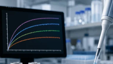
The affected enzymes spanned three classes: hydrolases, oxidoreductases, and transferases. For a cysteine protease, the Km decreased from 12.4 µM to 8.1 µM—a 35% increase in apparent affinity—while Vmax remained unchanged. For a glutathione S-transferase, the opposite occurred: Vmax rose by 40% without a shift in Km. Most striking was a dual-specificity phosphatase whose activity curve inverted: at low substrate concentrations, the COMU-synthesized peptide was hydrolyzed faster; at high concentrations, slower. The inversion was reproducible across three independent syntheses and two different labs.
The researchers ruled out trivial explanations. They swapped buffer lots, changed assay plates, and had a colleague from another institution repeat the measurements. The pattern held. They also tested peptides synthesized with a third reagent, HBTU, and found intermediate effects—suggesting that the phenomenon is not unique to one compound. The 11 affected enzymes shared no obvious sequence motif, but structural modeling hinted at a common thread: surface-exposed catalytic residues. The nine unaffected enzymes, including lysozyme and ribonuclease A, had deeply buried active sites, which may have been inaccessible to the trace contaminants.
These results raise a sobering possibility. Many published kinetic parameters for peptide substrates may have been influenced by the reagent used in synthesis. If labs routinely switch suppliers or reagents without documenting the change, the literature could contain systematic biases. The authors recommend that journals require reporting of synthesis reagents and lot numbers, much as antibody validation data is now expected.
Why the Other Nine Stayed Silent
Lysozyme, an enzyme that cleaves bacterial cell wall peptidoglycan, showed no change in activity regardless of which reagent was used. Ribonuclease A, which degrades RNA, was similarly unaffected. The nine insensitive enzymes included both soluble and membrane-associated proteins, with catalytic sites ranging from shallow grooves to deep pockets. Why did they resist? The team turned to molecular docking simulations, using the known structures of contaminant molecules and the enzymes.
The simulations suggested that HOAt and Oxyma preferentially bind to regions with exposed aromatic or positively charged residues near the active site. In the affected enzymes, these binding pockets were accessible; in the insensitive ones, steric hindrance or electrostatic repulsion prevented stable interactions. For example, the phosphatase that showed the inverted curve had a wide, solvent-accessible active site lined with arginine and tyrosine. Lysozyme’s active site, by contrast, is a deep cleft with a narrow entrance. The contaminant molecules simply could not reach the catalytic machinery.
This structural explanation is plausible but not yet proven. The team acknowledges that other factors—such as differences in peptide sequence or residual solvent—could contribute. They have initiated a broader screen with 50 enzymes to map the sensitivity patterns more systematically. If the correlation with active-site accessibility holds, it could allow researchers to predict which assays are at risk and adjust their controls accordingly.
Lessons for High-Throughput Screening
High-throughput screening (HTS) labs often purchase peptide substrates from commercial vendors or synthesize them in bulk. The reagent used in synthesis is rarely specified in the assay protocol. If a vendor switches from HATU to COMU, or from one lot to another, the change could introduce systematic errors that mimic inhibitor or activator effects. The cost of such errors is high: false positives waste follow-up resources, and false negatives may cause promising compounds to be discarded.
The study’s authors propose several practical measures. First, include a reagent blank control—a peptide synthesized without the target sequence—in every screening plate. Second, track reagent lot numbers and synthesis dates, and flag any changes in kinetic baselines. Third, when a new batch of peptide is introduced, re-run a small panel of control enzymes to detect shifts. These steps add perhaps 5–10% to the cost of screening, but the reliability gains could be substantial. As one of the authors put it, “We spend millions on compound libraries and automation. Spending a few thousand on controls seems like common sense.”
The finding also connects to broader concerns about reproducibility in biomedical research. A 2016 survey in Nature found that more than 70% of researchers had tried and failed to reproduce another lab’s experiments. Undocumented reagent changes may be one underappreciated source of that failure. The peptide synthesis field has long known that coupling reagents can leave traces; the new work shows these traces can propagate into functional assays. It is a cautionary tale that applies beyond peptides—to any biochemical assay where the substrate is chemically synthesized.
A Toolkit for Method Cross-Contamination Checks
The team has made its data and protocols openly available, hoping that other labs will adopt them. The core toolkit is straightforward: LC-MS analysis of final peptide batches to look for reagent-related peaks; spike-in experiments where known amounts of HOAt or Oxyma are added to pure peptides to calibrate the effect; and a standard panel of five enzymes (two hydrolases, two transferases, one phosphatase) that can serve as early-warning sensors. The panel is designed to be run on a plate reader in a single afternoon, requiring no specialized equipment beyond what most biochemistry labs already have.
Initial tests in two independent labs have reproduced the main findings. One lab reported that a different reagent, HBTU, produced intermediate effects, confirming that the phenomenon is not limited to HATU and COMU. Another lab found that the shifts diminished when peptides were subjected to additional purification steps, such as preparative HPLC with a longer gradient. This suggests that more rigorous purification can reduce but not eliminate the contamination. The team recommends that researchers who observe unusual kinetic behavior in peptide substrates consider the synthesis history as a possible cause.
The open-source data set includes raw kinetic traces for all 20 enzymes, along with structural models and docking scores. It is available on a public repository, and the authors encourage others to contribute their own results. Over time, a database of reagent effects could help the community identify problematic combinations and establish best practices. The goal is not to alarm, but to inform: reagents are not inert carriers; they leave molecular fingerprints that can shape the data we collect.
Broader Implications for Biochemical Assays
The phenomenon observed with HATU and COMU is likely not limited to peptide coupling reagents. Many chemical reagents used in substrate synthesis—including protecting groups, cleavage cocktails, and scavengers—can leave residual traces that persist through purification. For example, trifluoroacetic acid (TFA), commonly used in peptide cleavage, can remain as a salt and affect enzyme activity at low pH. Similarly, scavengers like triisopropylsilane (TIPS) or phenol may carry over and interact with hydrophobic enzyme pockets. The team’s findings suggest that any synthetic step could introduce artifacts, and the only way to rule them out is through rigorous controls.
One counter-argument is that the observed effects might be specific to the particular enzymes or peptide sequences used. The researchers addressed this by testing a diverse panel and finding that sensitivity correlated with active-site accessibility. However, they also note that not all peptides are equally prone to contamination; longer or more hydrophobic sequences may trap more residual reagent. This variability means that labs working with a narrow set of substrates may never encounter the problem—and may be unaware of its potential. The authors emphasize that their goal is not to cast doubt on all published data, but to encourage awareness.
Another trade-off is the cost of additional controls. For a small academic lab, running a reagent blank for every peptide may double the synthesis effort. The team suggests a tiered approach: for routine assays, a single blank per batch; for critical measurements (e.g., drug discovery leads), full spike-in controls. This balances rigor with practicality. The cost of ignoring the issue, however, could be higher—wasted follow-up on false hits or missed opportunities from false negatives.
The study also raises questions about commercial peptide suppliers. Many vendors guarantee high purity but do not disclose the reagents used. If a supplier changes its process without notice, customers may see unexplained shifts in assay performance. The authors recommend that researchers request reagent information from vendors and include it in their methods sections. This would create market pressure for transparency.
Looking ahead, the team plans to extend the screen to other coupling reagents, including newer ones like DIC/Oxyma and HBTU, to build a comprehensive map. They also aim to develop a predictive model based on contaminant-enzyme docking scores, which could flag risky combinations before experiments begin. Such a tool would be a valuable resource for the community.
The Takeaway: Reagents Are Not Inert Carriers
This study is a reminder that every step in an experimental workflow leaves a trace. The swap from HATU to COMU was a minor logistical decision, yet it reshaped 55% of the activity profiles in a panel of enzymes. The effect was not random; it followed structural logic and was reproducible across labs. But it was also invisible to standard quality checks, hiding in plain sight. For the field of enzymology, the implication is clear: we need to pay attention to the molecular history of our substrates.
The authors do not claim that all published kinetic data are suspect. Many studies use internally consistent reagent lots and would not be affected. But the finding underscores the value of transparency. Reporting the synthesis reagent, lot number, and purification method for peptide substrates should become routine, much as we now report antibody catalog numbers and cell line authentication. The cost is low; the benefit is a more robust literature.
The broader lesson extends to any field where synthetic molecules meet biological systems. As the boundaries between chemistry and biology blur, the artifacts of one discipline become the artifacts of the other. The reagent swap that changed 11 curves is not a crisis; it is a calibration. It tells us that our methods are more interconnected than we often assume, and that the quiet choices in the synthesis lab can echo loudly in the assay plate. Listening for those echoes may help us build science that is not only reproducible, but also aware of its own fingerprints.